

Diagnostic Tests
Written by Olivia Zoph
Doctor Know features an array of diagnostic tests drawn from real-world practice and cutting-edge research. Some of these tools have been in use for decades. Others are have just come to market. And many are still in research labs but promise to transform medicine in the coming decade.
Click on the headings below to see more information about each diagnostic test. Explore them all!
Biosignature Click for more info

When a virus or bacteria enters your body, your cells start an all-out war against the invaders. Your immune system has first lines of defense to try to keep the invaders off. In the meantime, they make special soldiers, called antibodies, to fight them.
 Antibodies are very specific to different types of bacteria and viruses. This means
that they will only attach to one type of bacteria or virus. Once they’ve attached to the invader, it is
a signal for other cells of the immune system to destroy it. It also signals your body to make more and
more of the antibodies specific to that invader. Doctors can use this knowledge when they are using
biosignature to see what type of infection a patient has.
Antibodies are very specific to different types of bacteria and viruses. This means
that they will only attach to one type of bacteria or virus. Once they’ve attached to the invader, it is
a signal for other cells of the immune system to destroy it. It also signals your body to make more and
more of the antibodies specific to that invader. Doctors can use this knowledge when they are using
biosignature to see what type of infection a patient has.
Body fluids such as blood, saliva and urine can be collected from sick patients. These fluids hold the antibodies that are key to fighting the infection. The body fluid is poured over a slide that holds millions of molecular sensors. The antibodies will only attach to molecules that have an attachment point similar to the invaders'. Then, the plate is gently washed so that only the attached antibodies remain. Next, a special process makes the antibodies glow. This results in a glowing pattern or “signature”. Different types of infections reveal different signatures. These signatures can be used to identify which invader is causing the infection.
Body Cam
 When you visit the
doctor, they do their best to figure out what is wrong with you. They look in your ears and up your nose
and have you open your mouth and say "ahhhhh." They may even feel special nodes in your neck to feel
their size. But for some illnesses, doctors actually need to peek inside of your body, a task that can
be pretty hard.
When you visit the
doctor, they do their best to figure out what is wrong with you. They look in your ears and up your nose
and have you open your mouth and say "ahhhhh." They may even feel special nodes in your neck to feel
their size. But for some illnesses, doctors actually need to peek inside of your body, a task that can
be pretty hard.
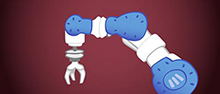
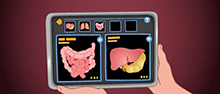
 Body Cam, also called endoscopy, is a tool that allows the doctor to look at the
inside of a patient's body. To do an endoscopy, a doctor inserts a camera into a natural opening
of the patient's body. This allows the doctor to look for signs of disease. Many parts of the body can
be seen with this tool, including the nose, ears, or parts of the digestive system. Doctors can also get
a better look at the respiratory system, urinary system, and female reproductive system. Other areas of
the body can be seen with cameras inserted into the body cavity through a small cut in the skin. Areas
of the body that can be seen this way include the inside of joints, the chest, and the abdominal cavity.
Body Cam, also called endoscopy, is a tool that allows the doctor to look at the
inside of a patient's body. To do an endoscopy, a doctor inserts a camera into a natural opening
of the patient's body. This allows the doctor to look for signs of disease. Many parts of the body can
be seen with this tool, including the nose, ears, or parts of the digestive system. Doctors can also get
a better look at the respiratory system, urinary system, and female reproductive system. Other areas of
the body can be seen with cameras inserted into the body cavity through a small cut in the skin. Areas
of the body that can be seen this way include the inside of joints, the chest, and the abdominal cavity.
In addition to being used to find problems, endoscopes are also used in surgeries. Surgeons insert a camera and surgical tools into the body cavity to complete some procedures. They do this through natural openings in the body or through small skin cuts. This way, they can see inside your body without having to make a larger cut. When endoscopy is used in the abdominal cavity it is called a laparoscopy. You or someone you know has probably had their appendix removed. The surgery to remove this organ is normally done laparoscopically.
Culture and Classify
 Some people say "what we
don't know can't hurt us," but in the body, that's quite a different story. Often, the things we can't
see are the things that bring the worst illnesses. Microbes are organisms that can only be seen with a
microscope. Despite their small size, they have the potential to make us very sick. Microbes can be
bacterial, fungal or viral. When doctors think that a microbe is making a person sick, they use culture
& classify to check. It is important to know exactly what type of microbe is causing the infection
so that it can be treated correctly.
Some people say "what we
don't know can't hurt us," but in the body, that's quite a different story. Often, the things we can't
see are the things that bring the worst illnesses. Microbes are organisms that can only be seen with a
microscope. Despite their small size, they have the potential to make us very sick. Microbes can be
bacterial, fungal or viral. When doctors think that a microbe is making a person sick, they use culture
& classify to check. It is important to know exactly what type of microbe is causing the infection
so that it can be treated correctly.
First, infected body fluid is collected from the patient. For example if someone has a sore throat, the doctor can take a sample of the saliva in the back of the throat to look for “bad microbes” that could be the cause. Once the sample is collected, it is sent to a lab for analysis.
 In order to make it easier to identify the “bad microbe”, lab scientists will try to
grow the microbe from the body fluid. In the lab, the microbe is given everything it needs to grow – the
right food, temperature, light and anything else it needs. The sample is often placed in a shallow dish
called a Petri dish. The Petri dish contains a gel of nutrients called agar that the microbe will feed
on while it grows. The Petri dish is often placed in an incubator that keeps the microbes at the perfect
temperature for them to grow. Over a few days, the microbes will replicate and grow. Once enough
microbes have grown in the Petri dish, scientists can perform tests to identify what type of microbe it
is. The lab scientists send a note to the doctor telling them which microbes they found. Once the doctor
knows what type of microbe was in the sample, they can choose the right treatment to fight it.
In order to make it easier to identify the “bad microbe”, lab scientists will try to
grow the microbe from the body fluid. In the lab, the microbe is given everything it needs to grow – the
right food, temperature, light and anything else it needs. The sample is often placed in a shallow dish
called a Petri dish. The Petri dish contains a gel of nutrients called agar that the microbe will feed
on while it grows. The Petri dish is often placed in an incubator that keeps the microbes at the perfect
temperature for them to grow. Over a few days, the microbes will replicate and grow. Once enough
microbes have grown in the Petri dish, scientists can perform tests to identify what type of microbe it
is. The lab scientists send a note to the doctor telling them which microbes they found. Once the doctor
knows what type of microbe was in the sample, they can choose the right treatment to fight it.
DNA Decoder
 DNA is a molecule in our
cells that tells the cell how to develop and function. The DNA molecule is a long double-helix (like a
twisted ladder) of nucleotides. There are four nucleotides in DNA. How the nucleotides line up in the
DNA molecule makes the instructions for the cells. Think of these nucleotides as letters in the alphabet
that make up the language of cells.
DNA is a molecule in our
cells that tells the cell how to develop and function. The DNA molecule is a long double-helix (like a
twisted ladder) of nucleotides. There are four nucleotides in DNA. How the nucleotides line up in the
DNA molecule makes the instructions for the cells. Think of these nucleotides as letters in the alphabet
that make up the language of cells.
 of nucleotides.") The order of these nucleotides is important. If we think about it in terms of words,
“read” and “dear” contain the same four letters, but they have very different meanings. Errors in our
DNA can lead to problems because the cells misread the instructions. Diseases caused by mutations in DNA
are called genetic diseases. Mutations in DNA can occur spontaneously or be passed down from our
parents.
The order of these nucleotides is important. If we think about it in terms of words,
“read” and “dear” contain the same four letters, but they have very different meanings. Errors in our
DNA can lead to problems because the cells misread the instructions. Diseases caused by mutations in DNA
are called genetic diseases. Mutations in DNA can occur spontaneously or be passed down from our
parents.
Sometimes clues from a patient’s symptoms lead a doctor to think that patient has a genetic disease. If so, they can use DNA decoder to check. DNA decoder takes a look at DNA to see if there are any changes or errors that could be causing disease. Blood, hair, skin or other tissues are used to sample a patient’s DNA. Scientists are then able to determine the order of the nucleotides in the DNA. This is called sequencing genes. Certain segments of DNA are compared to known segments with mutations that cause disease. We have a lot of DNA so we have to use supercomputers and "bioinformatics" to analyze the huge DNA sequence data files.
DNA decoder can help determine if a patient has a genetic disorder. Some genetic diseases are color blindness, hemophilia (a bleeding disorder), and albinism. Another genetic disease is alpha-1-antitrypsin deficiency, in which special proteins in your body break down certain body tissues. Genetic testing has allowed scientists and medical professionals to better understand certain diseases. This also allows for early diagnoses for patients.
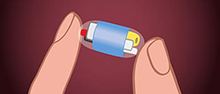
NanoDetectors
 When you test or explore
something, even a subject in class, it can be called probing. You are looking into a subject and
exploring, or probing, different parts of it. Doctors can also use special tools to probe parts of the
body. This helps them to treat or find out more about certain problems. For example, a dentist uses
special pointed tools to probe the edges of your teeth and gums to measure dental health. Deep wounds
can also be probed by doctors to let them see inside the wound and try to find whatever caused it.
When you test or explore
something, even a subject in class, it can be called probing. You are looking into a subject and
exploring, or probing, different parts of it. Doctors can also use special tools to probe parts of the
body. This helps them to treat or find out more about certain problems. For example, a dentist uses
special pointed tools to probe the edges of your teeth and gums to measure dental health. Deep wounds
can also be probed by doctors to let them see inside the wound and try to find whatever caused it.
. Image from Wikimedia via Crcatala") But a newer kind of probe that is much, much smaller can be used to treat other
problems. Very special nanoprobes are designed by scientists to explore your body and attach to specific
target molecules. The nanoprobes are injected into the patient's blood. In the blood they travel
throughout the body looking for the target molecules.
But a newer kind of probe that is much, much smaller can be used to treat other
problems. Very special nanoprobes are designed by scientists to explore your body and attach to specific
target molecules. The nanoprobes are injected into the patient's blood. In the blood they travel
throughout the body looking for the target molecules.
When the nanoprobes find the targeted molecule, they bind to it. After the nanoprobes are given enough time to find their targets, the patient is imaged to see where the nanoprobes have stopped. Doctors can see where they are because they are radioactive and release extra energy that can be picked up by a special machine called a PET scanner. The location of the nanoprobes can tell doctors if the patient has a disease.
 Nanoprobes are used in the diagnosis of Alzheimer's disease. Alzheimer's disease
mainly affects older people. It is a disorder that affects the brain and causes memory loss and
confusion. In patients with Alzheimer's disease the brain experiences a buildup of a protein called
Amyloid. If a doctor thinks a patient may have Alzheimer's disease, they can use nanodetectors to see if
there are amyloid plaques, or build-up, in the brain.
Nanoprobes are used in the diagnosis of Alzheimer's disease. Alzheimer's disease
mainly affects older people. It is a disorder that affects the brain and causes memory loss and
confusion. In patients with Alzheimer's disease the brain experiences a buildup of a protein called
Amyloid. If a doctor thinks a patient may have Alzheimer's disease, they can use nanodetectors to see if
there are amyloid plaques, or build-up, in the brain.
The patient is injected with special nanoprobes keyed in to find amyloid. If the patient has Alzheimer's disease, the PET scan will show the nanoprobes in the brain after they have attached to the amyloid plaques. If the patient does not have Alzheimer's disease the nanoprobes will be spread out in the body. This is because they will still be looking for molecules to which they can bind.
This technology is still being developed but has a lot of promise. While this tool can be helpful, it is only used in special situations because the nanoprobes are very, very expensive. Nanoprobe treatment also exposes the patient to radiation that can cause DNA damage.
Sample, Stain and Magnify
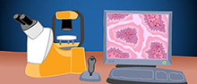 Small changes in our
body's cells can be a sign of disease. But cells are so tiny, how can doctors possibly see changes that
occur inside of cells? They can do this by using sample, stain, & magnify. First, a small
sample of tissue or cells is collected from the patient. This can be done with a cell scrape or a
biopsy. In cell scrape, doctors lightly scrape the tissue surface with a special brush or comb. This
gentle action will loosen and collect cells for the sample. In a biopsy, doctors gently cut away a small
section of tissue or group of cells. Biopsy is more invasive but it can show how cells interact with
their neighbors.
Small changes in our
body's cells can be a sign of disease. But cells are so tiny, how can doctors possibly see changes that
occur inside of cells? They can do this by using sample, stain, & magnify. First, a small
sample of tissue or cells is collected from the patient. This can be done with a cell scrape or a
biopsy. In cell scrape, doctors lightly scrape the tissue surface with a special brush or comb. This
gentle action will loosen and collect cells for the sample. In a biopsy, doctors gently cut away a small
section of tissue or group of cells. Biopsy is more invasive but it can show how cells interact with
their neighbors.
 After the doctor has collected the sample, it is sent to a lab for analysis. In order
to see the small structures inside the cell, the samples are stained with a colorful dye. Different
cells and different parts of cells take up dyes in different ways. Once the sample is dyed, scientists
or doctors can look at it under a microscope. The microscope magnifies the tiny cells. Based on what
parts are stained, the scientists and doctors can then identify the various parts of a cell and see what
is going on.
After the doctor has collected the sample, it is sent to a lab for analysis. In order
to see the small structures inside the cell, the samples are stained with a colorful dye. Different
cells and different parts of cells take up dyes in different ways. Once the sample is dyed, scientists
or doctors can look at it under a microscope. The microscope magnifies the tiny cells. Based on what
parts are stained, the scientists and doctors can then identify the various parts of a cell and see what
is going on.
Changes in the way the cells look can be a sign of an infection, cancer, or other disease. Cancer cells grow and replicate very quickly. They have a larger and darker nucleus that normal cells because the cancer cells contain more DNA. Because they’re growing so quickly, cancer cells also have a hard time staying organized and positioned in the right place. If a doctor sees disorganized cells with large, dark nuclei, it can be sign of cancerous cells. Doctors go through many years of training to be able to identify these and other small changes in cells.
Scan and Image
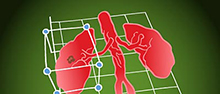
 The organs in our body
are protected by skin, muscles, and bones. This protection makes them difficult to evaluate for signs of
disease. Scan & Image helps doctors to see what’s going on inside the patient’s body. There are
several different imaging techniques doctors can use. Four common imaging techniques are X-ray, CAT
scan, MRI, and ultrasound. Each imaging technique has its own advantages and disadvantages and is used
in evaluating different tissues.
The organs in our body
are protected by skin, muscles, and bones. This protection makes them difficult to evaluate for signs of
disease. Scan & Image helps doctors to see what’s going on inside the patient’s body. There are
several different imaging techniques doctors can use. Four common imaging techniques are X-ray, CAT
scan, MRI, and ultrasound. Each imaging technique has its own advantages and disadvantages and is used
in evaluating different tissues.
 X-ray is
the most popular imaging technique. It uses electromagnetic x-ray radiation. Beams of x-ray radiation
are passed through the body. Different tissues absorb x-ray radiation differently. Dense tissue,
like bone, absorbs a lot of x-ray radiation, air absorbs very little and soft tissue is in between. The
amount of x-ray radiation that makes it to the other side is detected and measured and we get a picture
of the patient's insides.
X-ray is
the most popular imaging technique. It uses electromagnetic x-ray radiation. Beams of x-ray radiation
are passed through the body. Different tissues absorb x-ray radiation differently. Dense tissue,
like bone, absorbs a lot of x-ray radiation, air absorbs very little and soft tissue is in between. The
amount of x-ray radiation that makes it to the other side is detected and measured and we get a picture
of the patient's insides.
X-ray is good at looking for broken bones and it is quick and relatively inexpensive. It's only used if necessary though because in large amounts, X-ray radiation can cause mutations in the cell’s DNA and can increase the risk of cancer.
Screen Body Fluids
 Sweat can stink. But it
can also tell doctors about changes happening in your body. The makeup of many kinds of body fluids
(sweat, blood, urine, etc.) can change when a patient is sick or diseased. The amount and ratio of
cells, proteins, ions, enzymes, fats, sugars, and other elements can be measured in some of the fluids.
This information is compared to the values seen in a healthy adult. If the sample values are too high or
too low, it can be a sign of disease.
Sweat can stink. But it
can also tell doctors about changes happening in your body. The makeup of many kinds of body fluids
(sweat, blood, urine, etc.) can change when a patient is sick or diseased. The amount and ratio of
cells, proteins, ions, enzymes, fats, sugars, and other elements can be measured in some of the fluids.
This information is compared to the values seen in a healthy adult. If the sample values are too high or
too low, it can be a sign of disease.
 Doctors can screen body fluids to check these values. First, body fluid samples are
collected from the patient and sent to the lab. Lab scientists measure the different components of the
fluid and send a report of this information back to the doctor. These values help doctors to figure out
exactly what is wrong.
Doctors can screen body fluids to check these values. First, body fluid samples are
collected from the patient and sent to the lab. Lab scientists measure the different components of the
fluid and send a report of this information back to the doctor. These values help doctors to figure out
exactly what is wrong.
For example, doctors can order a complete blood count (CBC) on a sample of a patient’s blood to determine the number of different cells in the blood. Among other cells, lab scientists report how many white blood cells are in the sample. White blood cells are our infection-fighting cells. Having more white blood cells than normal can be a sign that the body is trying to fight off infection. Screening body fluids is an important tool but it only offers general clues, rather than specific answers.. With the help of clues from the patient’s history, physical exam, and other diagnostic tests, doctors can determine if the patient has an infection.
Stress and Measure
 If a chair is broken, you
may not realize it until you put weight on it. Some parts of the body are the same. Unless you put some
sort of stress on it, it's hard to tell how healthy it is.
If a chair is broken, you
may not realize it until you put weight on it. Some parts of the body are the same. Unless you put some
sort of stress on it, it's hard to tell how healthy it is.
Stress & Measure is a diagnostic tool doctors can use in order to see how well an organ is functioning when it is working hard. This tool is often used to test the heart and is referred to as a cardiac stress test. To test the heart when it is working hard the patient is placed on a treadmill (usually) that increases speed until the heart rate reaches a certain level.
 Doctors use an electrocardiogram (EKG) to evaluate the heart. The heart beats when a
wave of electricity passes through the cells. This electrical wave causes the cells to contract and
squeeze blood into the next chamber. Blood is pumped through the entire body by the action of the heart.
The wave of electricity can be measured with an EKG as it passes through the cells of the heart.
Doctors use an electrocardiogram (EKG) to evaluate the heart. The heart beats when a
wave of electricity passes through the cells. This electrical wave causes the cells to contract and
squeeze blood into the next chamber. Blood is pumped through the entire body by the action of the heart.
The wave of electricity can be measured with an EKG as it passes through the cells of the heart.
Additionally, doctors measure heart rate, breathing, blood pressure, and how the patient feels during the test. In a cardiac stress test, doctors can compare and measure the heart when the patient is resting and when it is working hard. Differences in the measurements can be a sign of disease.
Read more about: Doctor Know
You may need to edit author's name to meet the style formats, which are in most cases "Last name, First name."
https://askabiologist.asu.edu/diagnostic-tests
Bibliographic details:
- Article: Diagnostic Tests
- Author(s): Dr. Biology
- Publisher: Arizona State University School of Life Sciences Ask A Biologist
- Site name: ASU - Ask A Biologist
- Date published: 29 Apr, 2014
- Date accessed:
- Link: https://askabiologist.asu.edu/diagnostic-tests
APA Style
Dr. Biology. (Tue, 04/29/2014 - 08:43). Diagnostic Tests. ASU - Ask A Biologist. Retrieved from https://askabiologist.asu.edu/diagnostic-tests
American Psychological Association. For more info, see http://owl.english.purdue.edu/owl/resource/560/10/
Chicago Manual of Style
Dr. Biology. "Diagnostic Tests". ASU - Ask A Biologist. 29 Apr 2014. https://askabiologist.asu.edu/diagnostic-tests
Dr. Biology. "Diagnostic Tests". ASU - Ask A Biologist. 29 Apr 2014. ASU - Ask A Biologist, Web. https://askabiologist.asu.edu/diagnostic-tests
For more info, see http://owl.english.purdue.edu/owl/resource/717/04/
MLA 2017 Style
Modern Language Association, 7th Ed. For more info, see http://owl.english.purdue.edu/owl/resource/747/08/

Be Part of
Ask A Biologist
By volunteering, or simply sending us feedback on the site. Scientists, teachers, writers, illustrators, and translators are all important to the program. If you are interested in helping with the website we have a Volunteers page to get the process started.