


How Fast Do Embryos Grow?
Illustrated by: Sabine Deviche
show/hide words to know
Embryo: the egg after fertilization and before it has developed into a recognizable form.
Endoscope: a thin, long tube with a light and camera attached that is used to see inside the body...more
Fertilization: the process of uniting sperm and egg to begin embryo development... more
Fetus: an unborn animal that has started developing all of its body organs, but that is still in an early stage of development.
Menstrual Cycle: a monthly cycle that happens inside the female body. During the menstrual cycle, changing amounts of hormones result in changes in the ovaries and the tissue lining of the uterus. This prepares the body for possible pregnancy.
Organ: a specialized or distinct structure that is made from groups of tissues (e.g., heart, brain, etc.).
Ultrasound: a technique for making an image of what is inside the body......more
Uterus: or womb is a major female reproductive sex organ of most mammals including humans. It is within the uterus that the fetus develops... more
Have you ever looked at a picture of yourself from when you were little and wondered how you look so different now? Maybe your hair was lighter back then, or your nose was a slightly different shape. And of course, you’ve probably gotten a lot bigger.
It may seem like your body has changed a lot since then, but some of the most significant growth it’s gone through actually happened before you were even born.
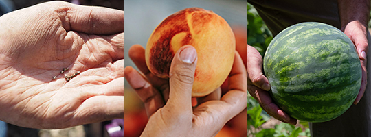
Over the course of a pregnancy, an embryo grows from being smaller than a poppy seed to as large as a small watermelon. Images via Wallpaper Flare, Michael Burrows via Pexels, and Mark Stebnicki via Pexels.
Over nine months of pregnancy, an embryo goes from being smaller than a poppy seed to the size of a watermelon. That’s a huge growth rate compared to what we go through after birth. Not only do embryos get bigger during that time, but they also go from being just a simple, single, cell to being the many cells that make up a human. All of a human’s features, organs, and body systems form when they're in the womb.
Let’s learn more about how embryos form, and how they grow throughout pregnancy.
Trimester 1
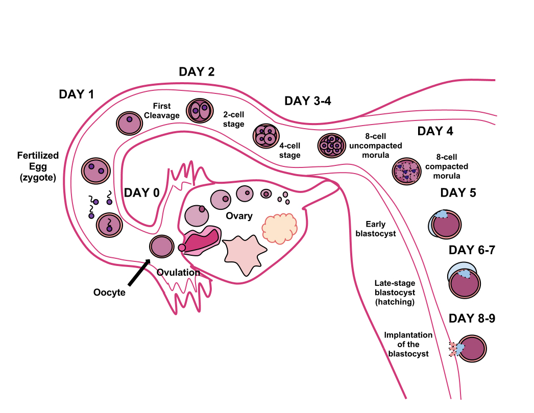
This image outlines the process of fertilization. Click the image for more detail.
The first trimester, or stage of pregnancy, includes the first 12 weeks. Doctors often start counting a pregnancy from the first day of a person’s last menstrual period. But the pregnancy really only starts after fertilization, when the sperm and egg meet to form a zygote. That typically occurs just over two weeks after the start of a pregnant person’s last menstrual period.
In the week after the zygote forms, it starts going through cell division. As that happens, the zygote becomes a blastocyst, or a hollow cluster of dividing cells. Around the end of that week, the blastocyst should implant, or attach, in the uterine wall. That’s where it will grow for the rest of pregnancy. About 10 to 12 days after fertilization, the blastocyst is considered an embryo.
When an embryo is three weeks old, a spine begins forming, as does the tissue that will become the heart. A spherical shape appears on the top part of the embryo that will become the head.
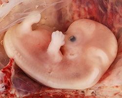
The image shows an embryo in the ninth week of pregnancy, or when it is seven weeks old. We can see the developing spine running through the embryo with the head on top. The black dot also indicates the early stages of the eye. Image of an embryo from an ectopic pregnancy by Ed Uthman via Wikimedia Commons.
Small buds that will grow into arms and legs appear on the embryo’s sides when it’s about four weeks old. Some early facial features, like eyes, also start to appear at that time.
Genitals and bones start to form when the embryo is five weeks old. By eight weeks, the fingers and toes are formed, though they have more growing to do. At the end of its eighth week, the embryo is considered a fetus.
The first trimester ends about 12 weeks after the start of a person’s last period, or when the fetus is about 10 weeks old. By that time, all of the fetus’s organs, limbs, bones, and muscles have started to form. It’s about the shape of a tiny human, but with a head that’s way too big for its body. About the size of a peach, the fetus still has a long way to go before all its parts are fully functional.
Trimester 2
The second trimester lasts from when the fetus is about 11 to 26 weeks old (weeks 13 – 28 of the pregnancy). All of the structures that formed in the first trimester continue to grow.

The embryo gets nutrients in different ways as it grows into a fetus. Click for more detail.
At as early as 12 weeks old, the external genitalia may show up on an ultrasound. The fetus’s skin starts to thicken and begins to act as a protective shield.
When the fetus is 14 weeks old, its ears have developed enough that it can hear its parent’s voice.
The fetus has fingernails by its 20th week, and lungs by its 22nd. By 25 weeks old, the fetus may be able to blink.
At the end of the second trimester, the fetus’s body parts are more proportional to what they will be at birth. But, it’s only about the size of a cauliflower, and will continue to grow and put on body fat to prepare for life outside the womb.
Trimester 3
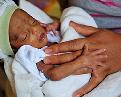
If a fetus is born before the 37th week of pregnancy, a doctor may call that a premature birth. That can come with certain health risks. Image by UNICEF Ethiopia via Flickr.
The third trimester is from the time the fetus is 27 weeks, or nearly seven months old, to the time it is born. Most are born around 38 weeks old, or 40 weeks after their parent’s last period. It’s the last stretch!
Have you ever heard a pregnant person say they can feel their baby kicking? That starts to happen when the fetus is about 27 weeks old, and is so big that it starts to feel cramped.
Thirty-two weeks in, most of the fetus’s organs are ready for birth, and it almost looks like a typical newborn. The two main organs still developing at that point are the lungs and the brain. The fetus’s bones are still developing, too, and will continue to for many years.
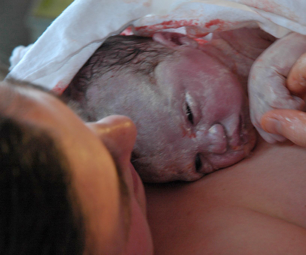
The white substance covering this newborn is called the vernix caseosa. Click for more detail.
After 38 weeks, the fetus is fully grown and ready to be born.
Most fetuses that develop the full 38 weeks weigh seven to nine pounds. They are about 20 inches, or 50 centimeters, long – about the size of a mini-watermelon. That’s a lot bigger than a zygote.
The fetus is usually born around 40 weeks after the start of the last menstrual period, ready to start life outside the womb.
Drawing Development
We haven’t always had ultrasounds to be able to check embryo growth inside the body. Previously, people had to study embryos in other ways.
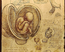
Here are da Vinci’s drawings of a fetus in the womb. You can see that da Vinci drew the position that a fetus takes during pregnancy. Image by Luc Viatour via Wikimedia Commons.
Have you heard of the Mona Lisa painting? The man who painted it, Leonardo Da Vinci, was also interested in science.
Sometime around 1510, Da Vinci began studying and drawing embryos and fetuses. His drawings were some of the first of their kind.
Even though he was making these drawings over 500 years ago, Da Vinci got a lot right. He was one of the first to show the human fetus in its proper position inside the womb. He also correctly guessed that the fetus grows faster in the womb than a child does after it’s born.
Creating the Carnegie Stages
Scientists continued to study embryos. But they didn’t have a good way to measure embryo development for hundreds of years.

This is the Carnegie Institute. For decades, researchers studied embryos there to create a way of sorting the stages of embryo growth. That led to the creation of the Carnegie Stages, which many scientists still use today. Image by AgnsoticPreachersKid via Wikimedia Commons.
Starting in the late 1800s, Franklin P. Mall, the first director of Carnegie Institution of Washington’s Department of Embryology, studied many embryos. He looked for patterns among the growth of embryos’ internal organs, external parts, and lengths. Based on those patterns, he tried to come up with a way to classify the stages of growth of embryos.
Mall died in 1917, but his project carried on for decades. In 1987, the Carnegie Institution published the staging system that resulted from all that work, called Carnegie Stages. The Carnegie Stages are still one of the most common ways of classifying the growth of human embryos in 2023.
Pictures for the Public
Though scientists had been studying embryos for decades, the general public had little idea what an embryo looked like. That is, until 1965, when Life Magazine published a story named “Drama of Life Before Birth.” Lennart Nilsson, a photojournalist, took photos of both embryos and fetuses for the story.
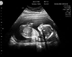
Nowadays, we can see the fetus easily with ultrasounds. Doctors use ultrasounds frequently throughout pregnancy to see the fetus. That helps a doctor to check for any problems in development. Image by Michael Fürstenberg via Flickr.
Many of Nilsson’s images were of fetuses that were removed from the womb for medical reasons. But, using tools such as an endoscope, Nilsson was also able to capture one of the first pictures of a living fetus inside a womb.
In 1965, Nilsson also published a popular book named “A Child is Born,” which had photos of growing embryos and facts about embryo development. Nilsson’s photos were some of the first to show the public, firsthand, how embryos grow.
Today, we now know a lot about what a growing embryo looks like. But, embryo development is an intricate process, and there are still many mysteries around how the steps of that process work. Many scientists continue to study embryos, and we are learning more about the process every year.
Image of violinist by Marco Verch via Flickr.
This Embryo Tale was edited by Risa Aria Schnebly and is based on the following Embryo Project Encyclopedia articles:
Cartwright, J. (2017, November 21). Countdown to Life: The Extraordinary Making of You (2015), by the British Broadcasting Corporation and The Open University. Embryo Project Encyclopedia. ISSN: 1940-5030 http://embryo.asu.edu/handle/10776/13016.
Conley, O. (2010, June 30). "Drama of Life Before Birth" (1965), by Life Magazine and Lennart Nilsson. Embryo Project Encyclopedia. ISSN: 1940-5030 http://embryo.asu.edu/handle/10776/2024.
Conley, O. (2010, June 24). Lennart Nilsson (1922- ). Embryo Project Encyclopedia. ISSN: 1940-5030 http://embryo.asu.edu/handle/10776/1995.
Gilson, H. (2008, August 19). Leonardo da Vinci's Embryological Annotations. Embryo Project Encyclopedia. ISSN: 1940-5030 http://embryo.asu.edu/handle/10776/1928.
Gilson, H. (2008, August 19). Leonardo da Vinci's Embryological Drawings of the Fetus. Embryo Project Encyclopedia ISSN: 1940-5030 http://embryo.asu.edu/handle/10776/1929.
Maayan, I. (2010, November 20). In the Womb (2005), by Toby Mcdonald and National Geographic Channel. Embryo Project Encyclopedia ISSN: 1940-5030 http://embryo.asu.edu/handle/10776/2308.
Wellner, K. (2009, July 17). Carnegie Stages. Embryo Project Encyclopedia ISSN: 1940-5030 http://embryo.asu.edu/handle/10776/1981.
Zhang, M. (2013-09-17). A Child Is Born (1965), by Lennart Nilsson. Embryo Project Encyclopedia. ISSN: 1940-5030 http://embryo.asu.edu/handle/10776/6274.
External Sources:
Artal-Mittelmark, R. (2022, September). Stages of Development of the Fetus. Merck Manual Consumer Version.https://www.merckmanuals.com/home/women-s-health-issues/normal-pregnancy/stages-of-development-of-the-fetus
Cleveland Clinic. (n.d.). Blastocyst. https://my.clevelandclinic.org/health/body/22889-blastocyst#:~:text=About%2010%20to%2012%20days,it%20then%20becomes%20a%20fetus.
Cleveland Clinic. (n.d.). Fetal Development. https://my.clevelandclinic.org/health/articles/7247-fetal-development-stages-of-growth#:~:text=The%20embryonic%20stage%20lasts%20from,eyes%2C%20mouth%20and%20limbs%20form.
Huffman, J. (2023, July 3). Pregnancy. Encyclopedia Britannica. https://www.britannica.com/science/pregnancy#ref76035
Parents Editors. (2023, May 9). How Big is My Baby This Week? Here’s a Baby Fruit Size Chart. Parents. https://www.parents.com/pregnancy/week-by-week/how-big-is-your-baby-this-week/
Singh, G., & Archana, G. First Initial. (2008). Unraveling the Mystery of Vernix Caseosa. Indian Journal of Dermatology, 53(2), 54–60.
UNICEF. (n.d.). How music affects your baby’s brain: Mini parenting master class. https://www.unicef.org/parenting/child-development/how-music-affects-you...
View Citation
Bibliographic details:
- Article: How Fast Do Embryos Grow?
- Author(s): Megha Pillai, Logan Hunt
- Publisher: Arizona State University School of Life Sciences Ask A Biologist
- Site name: ASU - Ask A Biologist
- Date published: August 9, 2023
- Date accessed: April 17, 2024
- Link: https://askabiologist.asu.edu/embryo-tales/embryo-growth
APA Style
Megha Pillai, Logan Hunt. (2023, August 09). How Fast Do Embryos Grow?. ASU - Ask A Biologist. Retrieved April 17, 2024 from https://askabiologist.asu.edu/embryo-tales/embryo-growth
Chicago Manual of Style
Megha Pillai, Logan Hunt. "How Fast Do Embryos Grow?". ASU - Ask A Biologist. 09 August, 2023. https://askabiologist.asu.edu/embryo-tales/embryo-growth
Megha Pillai, Logan Hunt. "How Fast Do Embryos Grow?". ASU - Ask A Biologist. 09 Aug 2023. ASU - Ask A Biologist, Web. 17 Apr 2024. https://askabiologist.asu.edu/embryo-tales/embryo-growth
MLA 2017 Style

Did you know that a fetus can hear music in the womb? Around the third trimester, a pregnant person’s fetus can hear music. Researchers say playing classical music or lullabies can soothe the fetus. They even say that music can help with brain growth before birth.
Image by Andrea Piacquadio via Pexels.
Be Part of
Ask A Biologist
By volunteering, or simply sending us feedback on the site. Scientists, teachers, writers, illustrators, and translators are all important to the program. If you are interested in helping with the website we have a Volunteers page to get the process started.