
Ectopic Pregnancy: An Unexpected Path
Written by: Logan Hunt
Illustrated by: Sabine Deviche
Illustrated by: Sabine Deviche
Culdocentesis: a medical procedure that uses a needle to remove fluid from a pouch behind the vagina...
... more
Embryo: the egg after fertilization and before it has developed into a recognizable form.
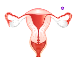
Fallopian tube: a structure inside female body that connects an ovary to the uterus.
Laparoscopy: a type of surgery in which a doctor makes a small incision on the lower stomach and inserts a small camera to see inside the body.
Laparotomy: a surgery that uses small cuts on the abdominal wall to gain access to the abdominal cavity. Using a camera to see inside the abdominal cavity is called laparoscopy...
... more
Placenta: a structure that forms in a pregnant person's body. It provides nutrients to the embryo while removing the waste from the embryo's blood...
... more
Uterus: or womb is a major female reproductive sex organ of most mammals including humans. It is within the uterus that the fetus develops... more
Kim runs into the living room to tell her partner Dan that she is pregnant. They both begin crying happy tears. This news brings a lot of emotions, as Kim and Dan have not had a healthy pregnancy yet.
It took eighteen months before Kim even became pregnant for the first time. After that, she was pregnant twice, but neither pregnancy lasted.
What went wrong before?
Both of Kim’s prior pregnancies were ectopic, so doctors had to remove the embryos. An ectopic pregnancy is when the embryo does not make it to the uterus to grow there like it would in a normal pregnancy.
The embryo instead grows in one of the fallopian tubes, which cannot support the growth of the embryo like a uterus can. As the embryo cannot survive anywhere but the uterus, Kim’s pregnancies both failed.
Let's look more closely at ectopic pregnancy and why it is so dangerous.
An Embryo’s Incomplete Journey
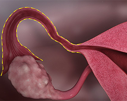
An ectopic pregnancy starts like a normal pregnancy—sperm from the male fertilizes the egg of a female, forming an embryo. The joining of the sperm and the egg always occurs in the fallopian tube. In a normal pregnancy, the embryo would then travel to the uterus to attach and grow for the rest of the pregnancy. But in an ectopic pregnancy the embryo does not make it to the uterus, and stays in the fallopian tube instead.
Ectopic pregnancies can be very dangerous for a pregnant person. Just as a huge truck can't fit through a tunnel made for small cars, the embryo has no room to grow in the fallopian tube.
Using the Uterus
In the uterus, the embryo has room to grow and the placenta can attach to the embryo. The placenta provides the embryo with nutrients from the pregnant person, so that the embryo can continue to grow throughout the pregnancy. Without those nutrients and in a small space, the embryo cannot survive.
How to Detect the Unexpected
Blood tests that look for hCG can also help determine if the pregnancy is ectopic. hCG is a hormone that the body produces when pregnant. A lower hCG level can be a sign of an ectopic pregnancy. That is because usually placental cells make the hormone hCG. When the embryo is not in the uterus, the placenta is much smaller, so it makes less of the hormone. Image via Pxfuel.
It is important to detect an ectopic pregnancy as soon as possible. The embryo gets bigger as the pregnancy goes on, which is risky for the pregnant person.
Usually, sharp pains in the lower back or stomach or abnormal bleeding are the first signs of an ectopic pregnancy. Nausea and fainting are also common early signs.
When an ectopic pregnancy ruptures, some fluids end up in areas where they shouldn’t be. The culdocentesis test checks the fluid behind the uterus in the abdomen of a pregnant person. When there is abnormal fluid present, it means the patient could have a ruptured ectopic pregnancy.
What Increases the Chances of Ectopic Pregnancy?
While ectopic pregnancies can occur at random, there are certain factors that increase the risk.
For example, Kim has endometriosis, which puts her at a higher risk of an ectopic pregnancy.
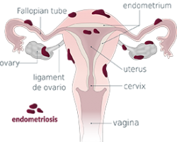
Endometriosis is the abnormal growth of the tissue that lines the uterus. The growths form outside of the uterus in places like the fallopian tubes. That means the embryo may attach to that abnormal tissue instead of the uterus. That causes an ectopic pregnancy.
When people cannot get pregnant on their own, they often turn to special technologies to help them. Those are procedures that aim to help the person get pregnant, but some also increase the risk of an ectopic pregnancy. If a fertilized egg is implanted into the fallopian tube, it may get stuck there and lead to an ectopic pregnancy.
Timely Treatment
To keep pregnant people safe and healthy, doctors need to treat ectopic pregnancies. Treatment depends on how far along the pregnancy is and what problems it causes. The goal of treatment is to protect the pregnant person’s health.
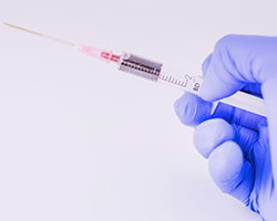
If caught early, a doctor can inject a medication called methotrexate to stop an ectopic pregnancy.
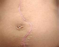
In other cases, surgery can remove the embryo. The surgery must be done carefully to not create too much scarring which could prevent the person from being able to become pregnant again. Laparotomy is a common surgery to achieve this, which removes the embryo and repairs any damaged tissue.
Understanding Ectopic Pregnancies
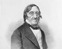
Scientists have been studying ectopic pregnancies for quite a long time. Regnier de Graaf was a Dutch physician who studied the body in the mid-seventeenth century. He was one of the first to recognize an ectopic pregnancy, which was long before people knew much about the egg.
Even though de Graaf did not know exactly what an egg was, he understood the importance of it moving to the right place. He noted that the most common place it stopped other than the uterus was the fallopian tube.
Over time, scientists have learned how to better explain ectopic pregnancy and they’ve worked on treatments for it.
Surgery Options for Ectopic Pregnancies
How do doctors remove the tiny embryo after an ectopic pregnancy has begun? The two main options for surgery are laparotomy and laparoscopy.
Laparotomy uses a large cut in the abdomen to perform the surgery. This method is used when the ectopic pregnancy is caught too late, resulting in a bigger embryo. The fallopian tube can also burst because of a growing embryo. If that happens, a laparotomy is necessary. In 1809, Ephraim McDowell carried out the first successful laparotomy.

Using the other option, a laparoscopy, a half-inch incision is made on the patient’s belly. The technique uses a tiny camera on the end of a small tube that goes into the patient through the incision. The doctors can then see inside the patient as they operate on the abdominal organs.
A laparoscopy leads to an easier recovery than a laparotomy, just like a paper cut usually heals more quickly than a scraped knee.
In 1901, Georg Kelling and Dimitri Von Ott separately created the surgery we now know as laparoscopy. In 1901, Kelling did some of the first laparoscopic procedures on dogs. In 1910, Hans Jacobaeus carried out the first laparoscopic procedure on a human.
With time things can always improve, right? In 1929, German gastroenterologist Heinz Kalk improved the lens used to see better inside the patient.
Modern Ectopic Pregnancies
We now have many tools to detect and treat ectopic pregnancies. Modern treatments such as medications and laparoscopy have also made it much safer to remove an embryo. So, while still dangerous, it is now easier to manage ectopic pregnancies than ever before.
Thumbnail image of ruptured fallopian tube by Hic et nunc via Wikimedia Commons.
Trumpet image by Marco Verch via Flickr.
This Embryo Tale was edited by Cole Nichols and is based on the following Embryo Project articles:
Cohmer, Sean, "2D Obstetric Ultrasound". Embryo Project Encyclopedia (2012-01-01). ISSN: 1940-5030 http://embryo.asu.edu/handle/10776/2328.
Conley, Olivia, "Laparoscopy". Embryo Project Encyclopedia (2010-06-19). ISSN: 1940-5030 http://embryo.asu.edu/handle/10776/1996.
Dupont, Ellen M., "Regnier de Graaf (1641-1673)". Embryo Project Encyclopedia (2008-09-30). ISSN: 1940-5030 http://embryo.asu.edu/handle/10776/1746.
Perez, Jovanna,, Santora, Emily, "Endometriosis". Embryo Project Encyclopedia (2021-06-21). ISSN: 1940-5030 http://embryo.asu.edu/handle/10776/13279.
Zhu, Tian, "Ectopic Pregnancy". Embryo Project Encyclopedia (2010-05-06). ISSN: 1940-5030 http://embryo.asu.edu/handle/10776/1934.
Read more about: Ectopic Pregnancy: An Unexpected Path
You may need to edit author's name to meet the style formats, which are in most cases "Last name, First name."
https://askabiologist.asu.edu/embryo-tales/ectopic-pregnancy
Bibliographic details:
- Article: Ectopic Pregnancy: An Unexpected Path
- Author(s): Logan Hunt
- Publisher: Arizona State University School of Life Sciences Ask A Biologist
- Site name: ASU - Ask A Biologist
- Date published:
- Date accessed:
- Link: https://askabiologist.asu.edu/embryo-tales/ectopic-pregnancy
APA Style
Logan Hunt. (). Ectopic Pregnancy: An Unexpected Path. ASU - Ask A Biologist. Retrieved from https://askabiologist.asu.edu/embryo-tales/ectopic-pregnancy
American Psychological Association. For more info, see http://owl.english.purdue.edu/owl/resource/560/10/
Chicago Manual of Style
Logan Hunt. "Ectopic Pregnancy: An Unexpected Path". ASU - Ask A Biologist. . https://askabiologist.asu.edu/embryo-tales/ectopic-pregnancy
Logan Hunt. "Ectopic Pregnancy: An Unexpected Path". ASU - Ask A Biologist. . ASU - Ask A Biologist, Web. https://askabiologist.asu.edu/embryo-tales/ectopic-pregnancy
For more info, see http://owl.english.purdue.edu/owl/resource/717/04/
MLA 2017 Style
Modern Language Association, 7th Ed. For more info, see http://owl.english.purdue.edu/owl/resource/747/08/

The fallopian tubes are named after Gabriele Falloppio, an Italian physician alive in the 1500s. He was one of the first people to describe the tubes, and he named them the "trumpets of the uterus."
Be Part of
Ask A Biologist
By volunteering, or simply sending us feedback on the site. Scientists, teachers, writers, illustrators, and translators are all important to the program. If you are interested in helping with the website we have a Volunteers page to get the process started.