

Hydraulic: caused by liquid under pressure.
Symmetry: equally balanced arrangement.
Sea Urchin Anatomy
One look at a sea urchin and you can see why they would be called sea hedgehogs. They have hard rounded shells covered with sharp movable spines. Urchins are part of the phylum Echinoderm and their name comes from Ancient Greek (echinos meaning "hedgehog" and derma meaning "skin"). There are more than 900 species of sea urchins and they come in a range of colors including purple, blue, brown, green, and red.
If you look inside a sea urchin you will see there are some organs that are familiar, like the esophagus and intestine. There are also some parts that are different than what you find in many other animals. The most unique organs are the ones that are part of the water system that acts like a hydraulic pump.
Looking Inside a Sea Urchin

| 1 | - | Genital plate | There are several plates surrounding the anus. Each plate has a single duct where the gametes (eggs or sperm) can be released into the water. |
| 2 | - | Gonopore | This is the opening at the end of the duct in the genital plate where eggs and sperm are released. |
| 3 | - | Anus | Notice the anus is located at the top of the urchin while the mouth is at the bottom. |
| 4 | - | Hard plate with madreporite | Water enters through this space into the water vascular system of the urchin. |
| 5 | - | Axial gland | This gland has been investigated by biologists for some time. It has been thought to be part of the immune system and part of the circulatory system. |
| 6 | - | Gonad | Organ that makes gametes (sperm or eggs). Sea urchin eggs have a golden color. |
| 7 | - | Intestine | Makes digestive enzymes and further breaks down food that came from the esophagus. |
| 8 | - | Ampullae | When the ampullae contract, the tube feet stretch as water moves into them. This allows the urchin to extend their tube feet. Muscles are used to contract the tube feet and push the water back into the ampullae. |
| 9 | - | Test | This is the exoskeleton, which is made of calcium carbonate. Our bones also have calcium carbonate in them to give them strength. |
| 10 | - | Radial canal | Ampullae branch off from either side of the radial canals. |
| 11 | - | Esophagus | The tube that runs through the center of Aristotle's Lantern and passes food to the intestine. |
| 12 | - | Aristotle’s lantern | The organ that chews and ingests food. |
| 13 | - | Teeth | These teeth act like jaws. There are five teeth in most sea urchins and they are part of the organ called Aristotle's lantern. |
| 14 | - | Mouth | Believe it or not, urchins have lips of soft tissue that also have small bony pieces embedded inside. |
| 15 | - | Nerve ring | Sea urchins can detect touch, chemicals, and light. |
| 16 | - | Ring canal | A muscular ring that circles the esophagus and is part of the water vascular system. |
| 17 | - | Test plates | Sections that form the test (exoskeleton). |
| 18 | - | Tube feet | Used for locomotion and to move loose food particles toward the mouth. |
| 19 | - | Spines | Used for defense and movement. If broken they can regenerate. |
Sea Urchin Water System
The water system of a sea urchin helps control its tube feet, which allow it to move and to grasp food particles. It works like a hydraulic system. Sea urchins aren't the only animals with a water system like this. Other echinoderms like sea stars also have a water system.

| 1 | - | Madreporite | A filtered opening that allows water into the water vascular system. |
| 2 | - | Aquifer | Water-filled area. |
| 3 | - | Ring canal | The ring canal circles the intestine and has five branches called radial canals. |
| 4 | - | Radial canal | There are five radial canals that supply water to the tube feet. |
| 5 | - | Ampulla | Part of the tube foot that expands when water is forced inside and contracted by using internal muscles. |
| 6 | - | Podia | Suction end found at the end of each tube foot. |
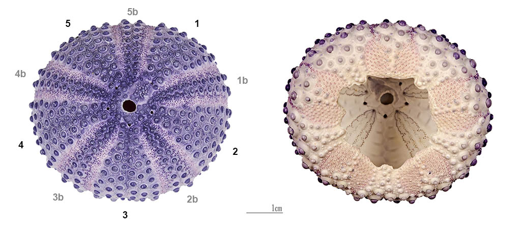
Sea Urchin Symmetry
You might not know it, but sea urchins have a fivefold symmetry just like sea stars. While the sea star's five arms make it easy to see its symmetry, it is not as obvious that the closely related sea urchin also has a fivefold symmetry.
Read more about: Sea Urchins Do Research
You may need to edit author's name to meet the style formats, which are in most cases "Last name, First name."
https://askabiologist.asu.edu/sea-urchin-anatomy
Bibliographic details:
- Article: Urchin Anatomy
- Author(s): Dr. Biology
- Publisher: Arizona State University School of Life Sciences Ask A Biologist
- Site name: ASU - Ask A Biologist
- Date published:
- Date accessed:
- Link: https://askabiologist.asu.edu/sea-urchin-anatomy
APA Style
Dr. Biology. (). Urchin Anatomy. ASU - Ask A Biologist. Retrieved from https://askabiologist.asu.edu/sea-urchin-anatomy
American Psychological Association. For more info, see http://owl.english.purdue.edu/owl/resource/560/10/
Chicago Manual of Style
Dr. Biology. "Urchin Anatomy". ASU - Ask A Biologist. . https://askabiologist.asu.edu/sea-urchin-anatomy
Dr. Biology. "Urchin Anatomy". ASU - Ask A Biologist. . ASU - Ask A Biologist, Web. https://askabiologist.asu.edu/sea-urchin-anatomy
For more info, see http://owl.english.purdue.edu/owl/resource/717/04/
MLA 2017 Style
Modern Language Association, 7th Ed. For more info, see http://owl.english.purdue.edu/owl/resource/747/08/

A sea urchin shell shows its fivefold symmetry. If you look carefully you can see the pattern.
Picture by Steve Jurvetson from Menlo Park, USA.
Be Part of
Ask A Biologist
By volunteering, or simply sending us feedback on the site. Scientists, teachers, writers, illustrators, and translators are all important to the program. If you are interested in helping with the website we have a Volunteers page to get the process started.