

Bubonic plague: bacterial disease carried by fleas of infected Old English rats. At its worst, it killed two million people a year. Those that caught the disease had a 90% chance of dying from it.
Great Plague of London: there have been several large outbreaks of the bubonic plague in Europe beginning in 1347. In 1665 London fell victim to the plague that has since been called the Great Plague of London... more
Microscope: an instrument used to see objects or parts of objects, which are too small to be seen with only our eyes... more
Robert Hooke (28 July 1635 – 3 March 1703)
The year was 1665. A book of illustrations called Micrographia has just been published by the English natural philosopher, Robert Hooke. The camera had not yet been invented so illustrations were common for books and other publications. What was uncommon about Micrographia was that it was one of the first time drawings of the microscopic world had been published.
Within the publication more than 30 detailed illustrations appeared including the famous one from cork that provided the first documentation of a single cell. Hooke also examined hair under a microscope and made a note that some of the hairs were split at the ends. This is possibly the first notation of split ends.
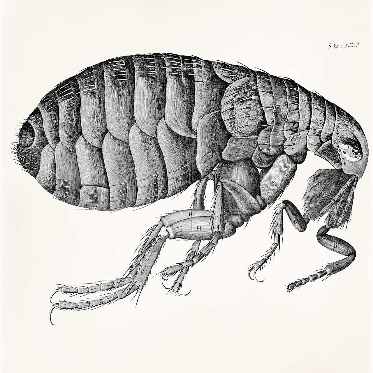
Examples of Hooke's detailed drawings can be seen in the illustration of a cork and a flea below. It was in his description of cork that he first used the term "cell" even though he did not know how important his discovery would become. The cell wasn't really understood until 1839 when scientists began to discover its importance.
Why Call it a Cell?
Hooke's drawings show the detailed shape and structure of a thinly sliced piece of cork. When it came time to name these chambers he used the word 'cell' to describe them, because they reminded him of the bare wall rooms where monks lived. These rooms were called cells.
Gallery of Images from Micrographia
There were no cameras when Robert Hooke first explored the tiny world with his microscope. To bring these images to life and share them with the world, he had to draw what he saw using his new instrument. Here are a few of the amazing drawings he made and published in 1665.

|
|
|
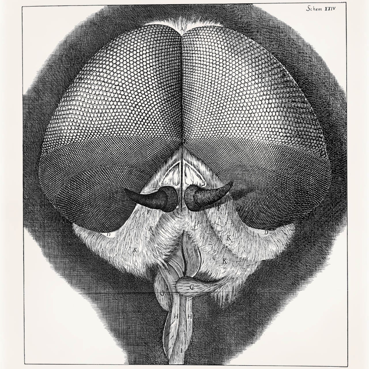
| Thin slice of cork. Click on the image to enlarge. | Drone-fly head. Click on the image to enlarge. | |
|
|
|
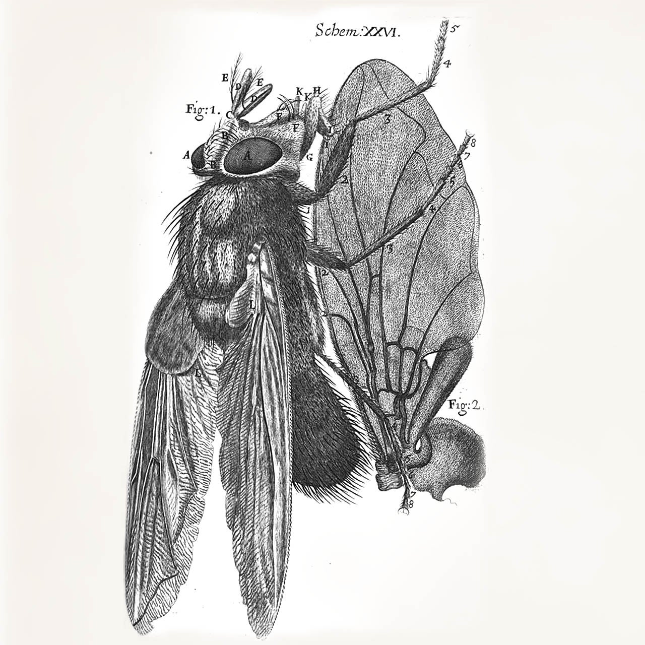
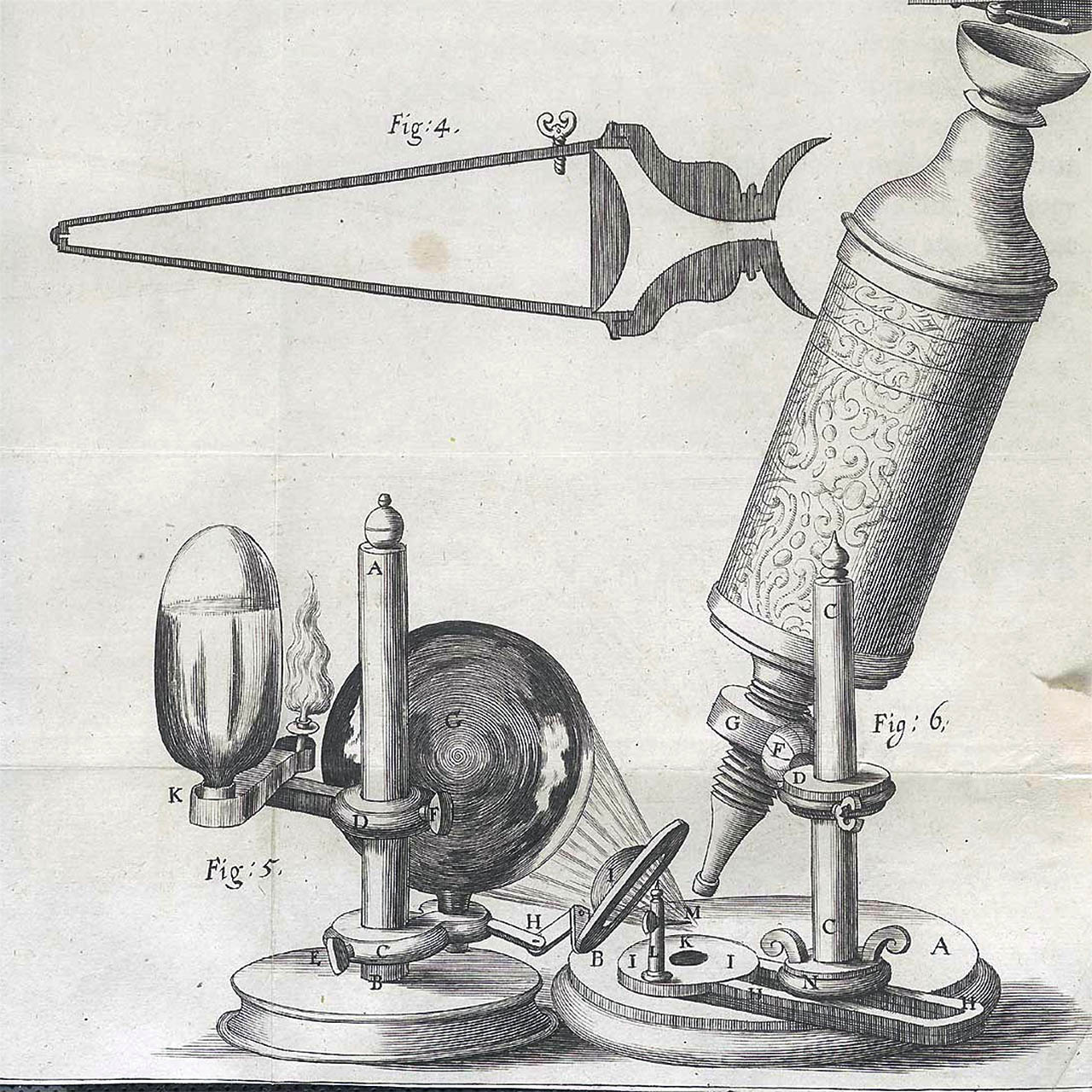
| Blue fly. Click on the image to enlarge. | A drawing of his microscope. Click on the image to enlarge. | |
|
|
|
|
As it turned out, the flea (Ceratophyllus fasciatus) in this illustration was the carrier of the Bubonic Plague that was sweeping through Europe at the time. However, Hooke did not know this. Click on the image to enlarge. |
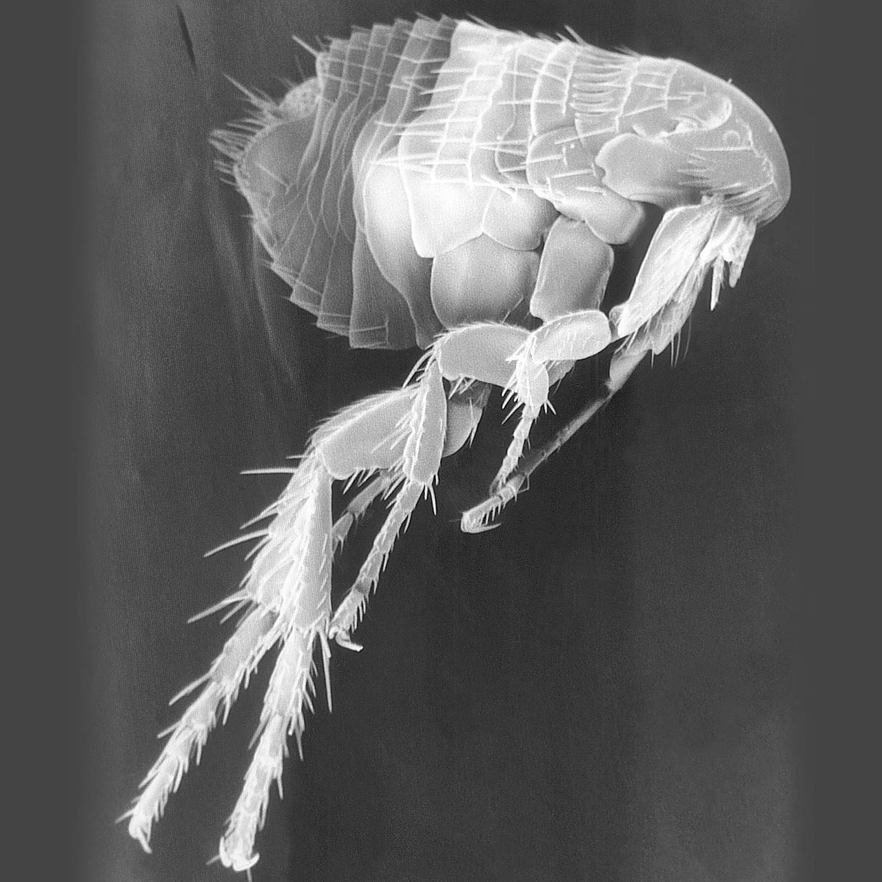
A flea imaged using a scanning electron microscope. Notice how much detail Hooke was able to see with his early microscope. Click on the image to enlarge. |
|

|
|
|
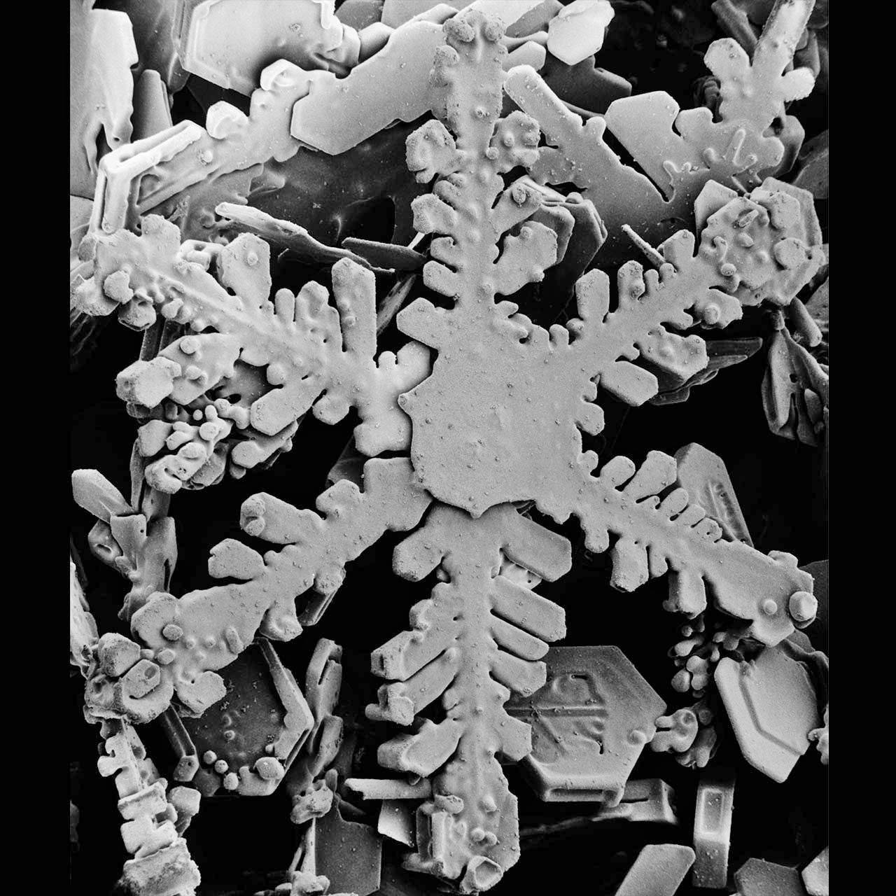
| Hooke also looked at things other than insects and plants. Here he has drawn several snow flakes. Click on the image to enlarge. | Scanning electron microscope image of a snowflake. Hooke was very accurate even with his early microscope. Click on the image to enlarge. |
Additional places to explore:
Read Micrographia and view all the images at Project Gutenberg.
References:
Historical Anatomies on the Web (NIH).
Scanning Electron Image of the flea from the Center for Disease Control (CDC)
Image of the scanning electron microscope snowflake from Beltsville Electron Microscopy Unit, part of the USDA.
Additional images from Wikimedia Commons.
Read more about: Building Blocks of Life
You may need to edit author's name to meet the style formats, which are in most cases "Last name, First name."
https://askabiologist.asu.edu/robert-hooke
Bibliographic details:
- Article: Robert Hooke
- Author(s): Dr. Biology
- Publisher: Arizona State University School of Life Sciences Ask A Biologist
- Site name: ASU - Ask A Biologist
- Date published:
- Date accessed:
- Link: https://askabiologist.asu.edu/robert-hooke
APA Style
Dr. Biology. (). Robert Hooke. ASU - Ask A Biologist. Retrieved from https://askabiologist.asu.edu/robert-hooke
American Psychological Association. For more info, see http://owl.english.purdue.edu/owl/resource/560/10/
Chicago Manual of Style
Dr. Biology. "Robert Hooke". ASU - Ask A Biologist. . https://askabiologist.asu.edu/robert-hooke
Dr. Biology. "Robert Hooke". ASU - Ask A Biologist. . ASU - Ask A Biologist, Web. https://askabiologist.asu.edu/robert-hooke
For more info, see http://owl.english.purdue.edu/owl/resource/717/04/
MLA 2017 Style
Modern Language Association, 7th Ed. For more info, see http://owl.english.purdue.edu/owl/resource/747/08/

Portrait of Robert Hooke. This is an artist's impression of Robert Hooke. It was created from the descriptions by his colleagues Aubrey and Waller. Image via Wikimedia. Painting by Rita Greer.
Be Part of
Ask A Biologist
By volunteering, or simply sending us feedback on the site. Scientists, teachers, writers, illustrators, and translators are all important to the program. If you are interested in helping with the website we have a Volunteers page to get the process started.