Protein Art... and What Proteins Really Look Like
Individual proteins are so small that you can’t actually see them, not even with most microscopes. So when you see a “picture” of a protein, you are really looking at a drawing or computer model of the protein’s structure.
Have you ever seen a model of our solar system? Maybe you made one for your science class out of Styrofoam balls, or maybe you've seen a really big model at a museum. These models help us imagine where the sun and planets are compared to the earth - even if the model is very simple. More detailed models can help us predict the motion of the planets around the sun. In the same way, different kinds of protein models can be used to visualize the structure of the protein in different ways.
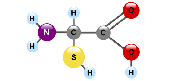
Breaking the Code
The way the amino acids (and other molecules) are drawn is like a secret code. Here’s how to break the code:
- Every "C" is a carbon atom
- Every "O" is an oxygen atom
- Every "N" is a nitrogen atom
- Every "S" is a sulfur atom
- Every "H" is a hydrogen atom
- Every "Se" is a selenium atom (Can you find the one amino acid that has selenium?)
- The hydrogen atoms that are attached directly to carbon atoms are sometimes not shown (think of them as invisible ninja hydrogens)
Sometimes to save space and time scientists will leave out the colored balls. This is mostly for the "H"s for hydrogen, and all the "C"s for carbon. If you see a model like this, you can still decode it. The carbon atoms are at the point of every angle in the line and at the end of any line that doesn't end in another letter.
Drawing amino acids in these ways helps us think about what shape the amino acids take and helps us predict how they will behave.
Alphabet Soup
So they don’t have to write the full name every time, scientists have abbreviations for the names of the amino acids. There are two ways – a three letter abbreviation and a one letter abbreviation. For example, arginine can be written as “Arg” or just as “R”. If you were a pirate, I bet your favorite amino acid would be arrrrrrginine. There has to be a standard code so nobody gets confused, especially because some of the amino acid names sound alike. For example, E is for glutamic acid and Q is for glutamine.
Let’s Explore Protein Structure
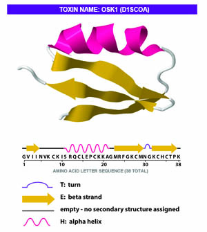
Scientists have many different ways to draw and look at protein shapes and structure. Depending on what they are interested in looking at they will pick different ways to draw and display the protein. Looking at the diagram on the right helps us see how the amino acid chain twists and bends to form the final protein shape, but is this really what the protein looks like?
The answer is…not really.
The Many Faces of Molecules
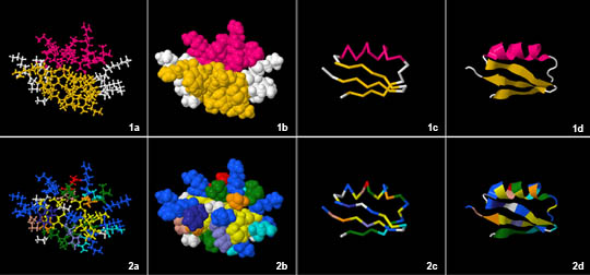
Scientists have developed many ways to show how molecules look, what they are made of, and how they might work. They call these drawings "models." Here are eight different models of the same OSK1 toxin protein that we just looked at. This is just one type of molecule found in scorpion venom, but it can have many different looks depending on what scientists are studying.
Color Coded Models
All of the models in the top row are color coded to show the different secondary structures. Pink is for spiral (helix) and yellow is for sheet. The bottom row is color coded by the different amino acids. For example, glutamic acid (E) is shown in white. You can see it on the bottom left “corner” of the protein model in all of the images on the bottom row. Now look back at the first model of OSK1 toxin that we saw. Can you find the end of the protein chain that starts with the amino acid glutamic acid (E)?
Why Are There So Many Ways to Draw Proteins?
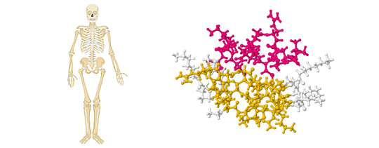
There are different kinds of images for different purposes. For example, here are some artistic ways to represent people and proteins.
Bare Bones
This skeleton is more like the ball and stick model (right) and 1a and 2a in our gallery of images above. Is it correct? Yes, it has all of the correct bones shown as well as how they connect to each other. Does it show you what an actual, living person looks like? Not exactly. This is how a ball and stick model of a protein works. It shows you where the important atoms are (balls) and how they connect to each other (sticks), but it doesn't give you a very good idea of the where the spirals (helices) and sheets are or the full shape of the protein.
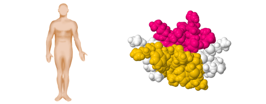
Fleshed Out
This drawing of a person shows the full human form. This is more like the space filling model (right) and 1b and 2b in our gallery of images above. This is the best way to imagine what the actual 3-D shape of the protein is like. Although there is a great amount of detail in a space-filling model, the image is so dense that it can make it difficult to tell what kind of structure the protein has (especially on the inside).

Stick Figure Art
Sometimes even a skeleton is too much information. If you just want the bare minimum of the protein structure, you can just show the backbone (right) like in 1c and 2c in our gallery of images above. This type of model ignores the structure of the individual amino acids and just shows the backbone of the protein (where the amino acids are hooked together).

Not Just For the Comics
In a comic strip, you just need the basics of arms, legs, and head to visualize what the character is doing. This simplification lets you focus on the most important parts, like the character's motion or emotions. For the ribbon diagram (right) and 1d and 2d in our gallery of images above, parts of the proteins' structures are minimized so that in each you can focus on the location of the one spiral (helix) and how the three parts of the sheet line up.
Addition images from Wikimedia.
Protein illustrations generated using Protein Data Bank (PDB) Jmol visualizer tool.
Read more about: Venom!
Bibliographic details:
- Article: Protein Art
- Author(s): Dr. Biology
- Publisher: Arizona State University School of Life Sciences Ask A Biologist
- Site name: ASU - Ask A Biologist
- Date published:
- Date accessed:
- Link: https://askabiologist.asu.edu/venom/protein-art
APA Style
Dr. Biology. (). Protein Art. ASU - Ask A Biologist. Retrieved from https://askabiologist.asu.edu/venom/protein-art
Chicago Manual of Style
Dr. Biology. "Protein Art". ASU - Ask A Biologist. . https://askabiologist.asu.edu/venom/protein-art
Dr. Biology. "Protein Art". ASU - Ask A Biologist. . ASU - Ask A Biologist, Web. https://askabiologist.asu.edu/venom/protein-art
MLA 2017 Style
Be Part of
Ask A Biologist
By volunteering, or simply sending us feedback on the site. Scientists, teachers, writers, illustrators, and translators are all important to the program. If you are interested in helping with the website we have a Volunteers page to get the process started.


