


show/hide words to know
Amino acid: molecules that contain carbon, oxygen, hydrogen, and nitrogen. These are the building blocks of protein......more
Diffraction: the ways in which waves act when they encounter an obstacle. In traditional physics, waves bend around the obstacle and they have specific behavior patterns after they pass an object... more
Humerus: the upper bone of the arm... more
Radiation: in physics, the way energetic particles or waves move through a medium (such as water or air) or space.
Symmetry: equally balanced arrangement.
X-ray: a photograph taken of the inside of the body using a special type of light... more
A Quick Class in X-ray Crystallography

X-ray of a right humerus bone including elbow joint and a portion of the shoulder joint. It shows a badly broken humerus from a wrestling match.
X-ray Diffraction
Have you – or someone you know – ever had a broken leg, arm or finger? In order to see if it was really broken, the doctor made a picture first. And not just an ordinary picture with a phone or digital camera, because with such a picture you can only see the limb from the outside. If you want to see if the bone inside the limb is damaged or broken, you have to be able to look right through your skin and flesh. X-ray photos can do this.
Radiation X
X-rays got their name from one of the first scientists who studied them: Wilhelm Röntgen. He did not know what type of radiation this was, so he called it “X”. Wilhelm Röntgen made great contributions to his field, and to acknowledge this many people named the rays after Wilhelm himself. They called it ‘Röntgen-rays’. In some languages this is still the commonly used name. However, more and more English speaking people started to use ‘X-ray’ and today this is the most used term.
Light Comes in Waves
What are X-rays? In order to understand this, let us first think of how we see the world around us. What we see with our eyes is all visible light. Light can have many different colors and that is why we see green leaves, red bricks and yellow snow. Why do we see these colors?
Already a long time ago scientists discovered that you can think of light as a very small wave. Just like you see wave of water on a lake or in the ocean. Some of these waves are a bit shorter and others are a bit longer. As it turns out, these differences in wavelength actually define the colour of the light. Waves of red light are for instance longer than waves of blue light.
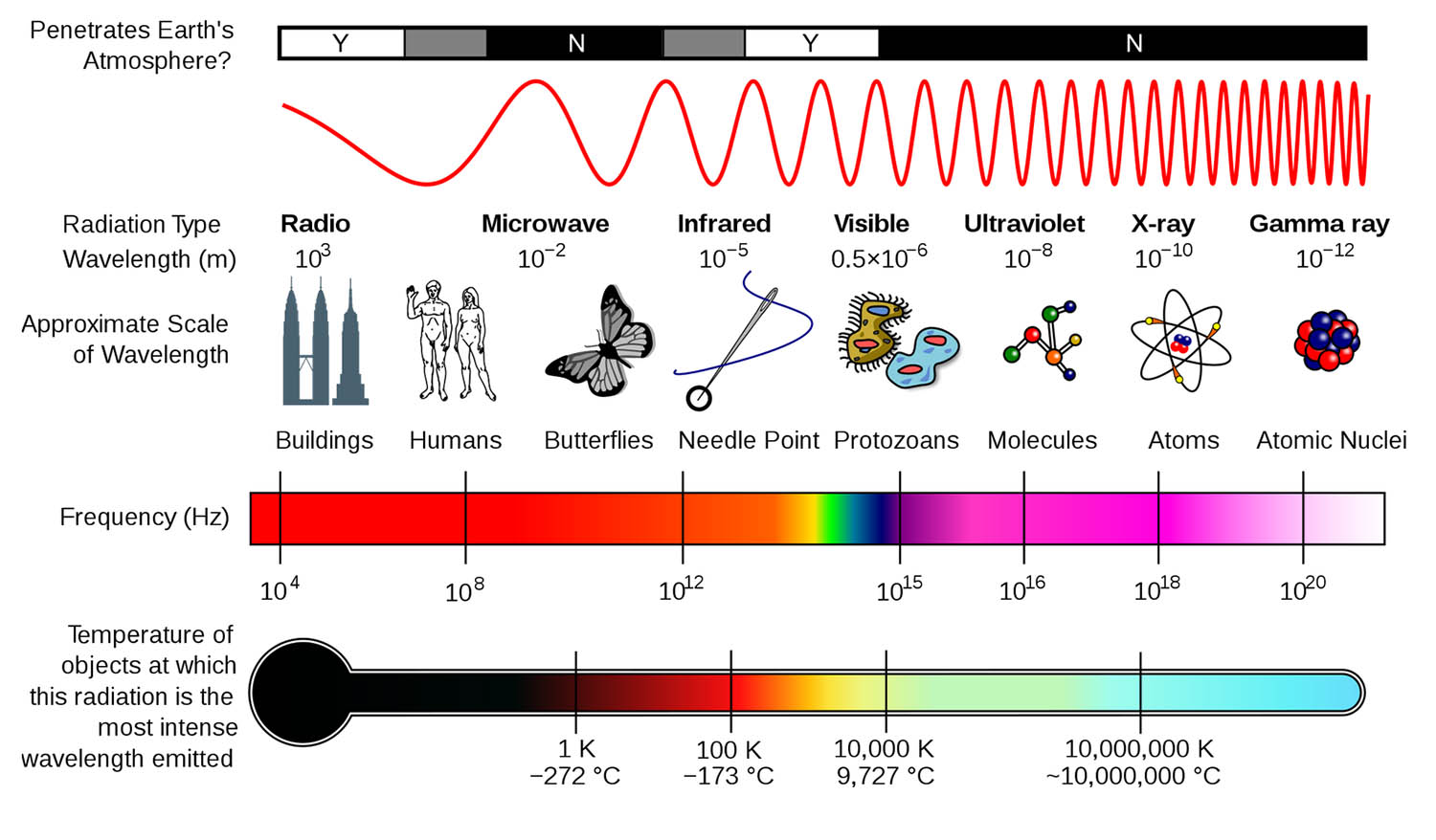
Types of light waves, their relative wavelength and the smallest object they can image. Click to see larger version.
X-rays, a Different Way of Looking
X-rays can also be seen as waves, but they are much shorter than light-waves. On top of this, X-rays can go right through some materials. Because of their special properties, we can use X-rays to see things that we cannot see with light. We cannot directly see what the X-rays show us with our eyes. Instead there are special cameras and detectors that can. These cameras can see X-rays and turn what they see into an image we can see. Using X-rays we can see right through the skin and tissue and look at a bone. Because X-ray waves are so small, they can also be used to look at very small things, like atoms inside proteins.
Diffraction
If waves go through a small opening or round a small object they bend around it and change direction. When this happens, scientists say the waves “diffract". This also happens to water waves. Sometimes waves in shallow water hit a rock that is about the same size as their wavelength. If this happens they bend around it and the rock appears to send new waves in all directions. X-ray waves can do the same, but only if they hit something that is about as small as they are, such as atoms.
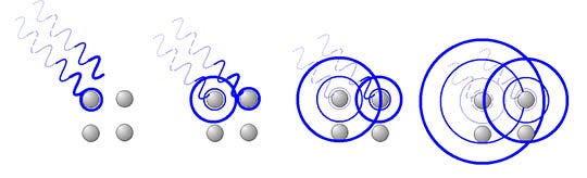
Illustration showing how X-rays interact with the atoms in a crystal.
X-ray Diffraction
Scientists use X-rays to find out what proteins look like in 3D. They do this by shooting a beam of X-rays at a protein crystal. If the beam hits the crystal, something very special happens. All atoms inside the crystal start to send out waves with the same wavelength, but then in all directions. This is comparable to what happens if water waves hit a rock as we described earlier. These waves that are sent out hit a plate, called a detector. This detector can see the X-rays and transforms what it sees into an image like one you see on the below right.
What’s in an Image?
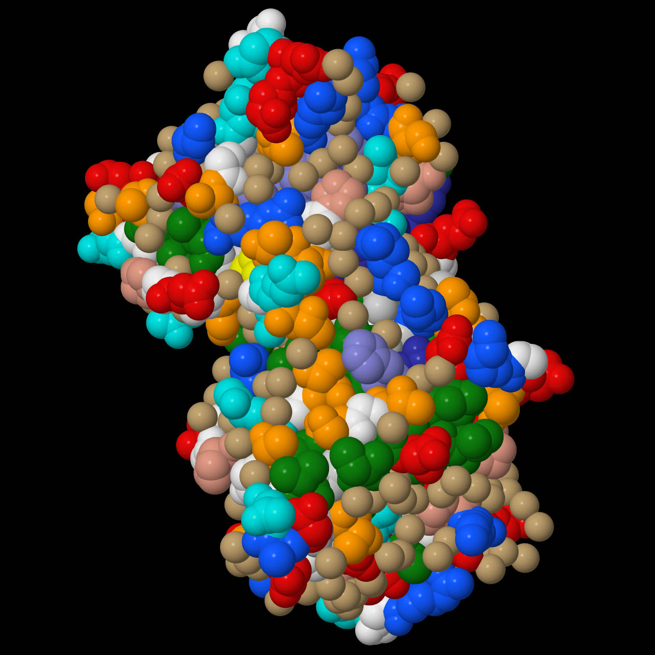
Photographic X-ray diffraction image from a bovine Cu,Zn superoxide dismutase crystal. Click on the image to get an idea of what it looks like in three dimensions.
Take a close look at the image to the right. Can you see the 3D shape of a protein in it? Scientists can’t either. But scientists know very well how X-rays behave if they hit an atom. With this knowledge they have made computer software that helps them to build a 3D model of the protein.
Scientists can obtain some information from the diffraction image directly. They can see if the protein is symmetrical. That is why Watson and Crick corrected their assumption when they saw the excellent Photo 51 from Rosalind Franklin. This photo clearly showed that DNA has a two-fold symmetry instead of a threefold. This is what lead them to discover that DNA has a double helical shape. Until then many scientists thought is was a triple helix.
Combining Imaging Tools to Get the Whole Picture
Scientists continue to study the shapes of molecules using X-ray
crystallography and other X-ray diffraction techniques. In many cases the molecules they want to study are large, or they even want to study a whole complex that consists of many proteins. For large objects, there is not one imaging technique that can gather the whole shape and at the same time the tiny details scientists want to see. In these cases, scientists use several imaging tools to collect information that is then pieced together. It is like building a very tiny jigsaw puzzle. Electron microscopes are one of the tools that are often used with X-ray diffraction to build 3D models of large proteins, DNA, RNA and viruses.
References:
Jason Dicker. Physics Help. Diffraction, Retrieved August 2012 from http://www.launc.tased.edu.au/online/sciences/physics/diffrac.html
Wikipedia, X-ray crystallography. Retrieved May 2012 from https://en.wikipedia.org/wiki/X-ray_crystallography,
Images from Wikimedia and Research Collaboratory for Structural Bioinformatics (RCSB).
View Citation
Bibliographic details:
- Article: A Quick Class in X-ray Crystallography
- Author(s): Martine Oudenhoven
- Publisher: Arizona State University School of Life Sciences Ask A Biologist
- Site name: ASU - Ask A Biologist
- Date published: August 19, 2012
- Date accessed: July 23, 2024
- Link: https://askabiologist.asu.edu/quick-class-x-ray-crystallography
APA Style
Martine Oudenhoven. (2012, August 19). A Quick Class in X-ray Crystallography. ASU - Ask A Biologist. Retrieved July 23, 2024 from https://askabiologist.asu.edu/quick-class-x-ray-crystallography
Chicago Manual of Style
Martine Oudenhoven. "A Quick Class in X-ray Crystallography". ASU - Ask A Biologist. 19 August, 2012. https://askabiologist.asu.edu/quick-class-x-ray-crystallography
Martine Oudenhoven. "A Quick Class in X-ray Crystallography". ASU - Ask A Biologist. 19 Aug 2012. ASU - Ask A Biologist, Web. 23 Jul 2024. https://askabiologist.asu.edu/quick-class-x-ray-crystallography
MLA 2017 Style
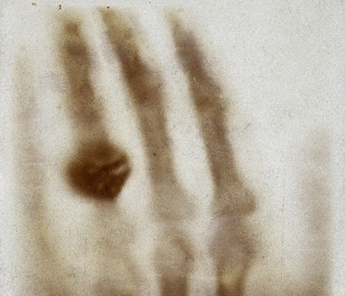
A print of one of the first X-rays by Wilhelm Röntgen (1845–1923) of the left hand of his wife Anna Bertha Ludwig.

Puzzles
Be Part of
Ask A Biologist
By volunteering, or simply sending us feedback on the site. Scientists, teachers, writers, illustrators, and translators are all important to the program. If you are interested in helping with the website we have a Volunteers page to get the process started.